Trinity Tales

Every pet has a story, and at Trinity Veterinary Hospital, we’re honored to be part of so many remarkable journeys. From life-saving surgeries to unexpected diagnoses, our team is committed to providing exceptional care, no matter the challenge. These tales of resilience, hope, and healing celebrate the special bond between pets and their families—and the extraordinary outcomes we’ve achieved together.
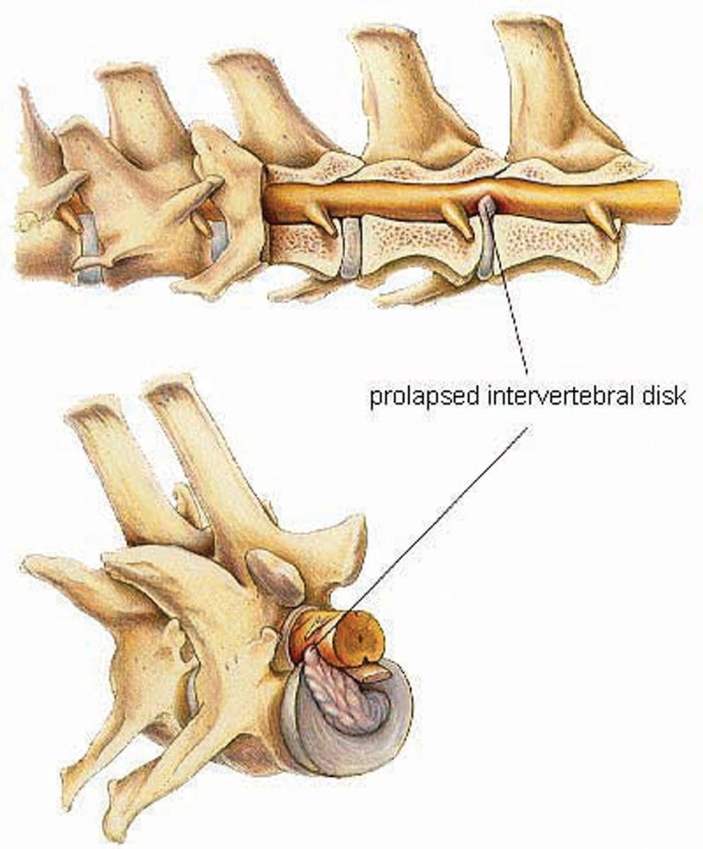
 Dachshunds are well-known for their elongated spines, which predispose them to intervertebral disc disease (IVDD). IVDD occurs when the shock-absorbing discs between the vertebrae progressively harden, losing their ability to cushion the vertebrae properly. Hardened discs often bulge and compress the spinal cord, disrupting nerve signals that control essential functions like bladder and bowel movements. In some cases, a minor jump or poor landing can cause a hardened disc to rupture, exerting pressure on the spinal cord, resulting in pain, nerve damage, or even paralysis.
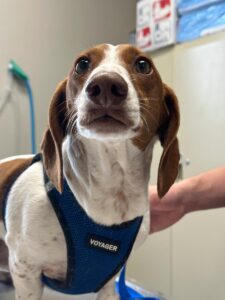
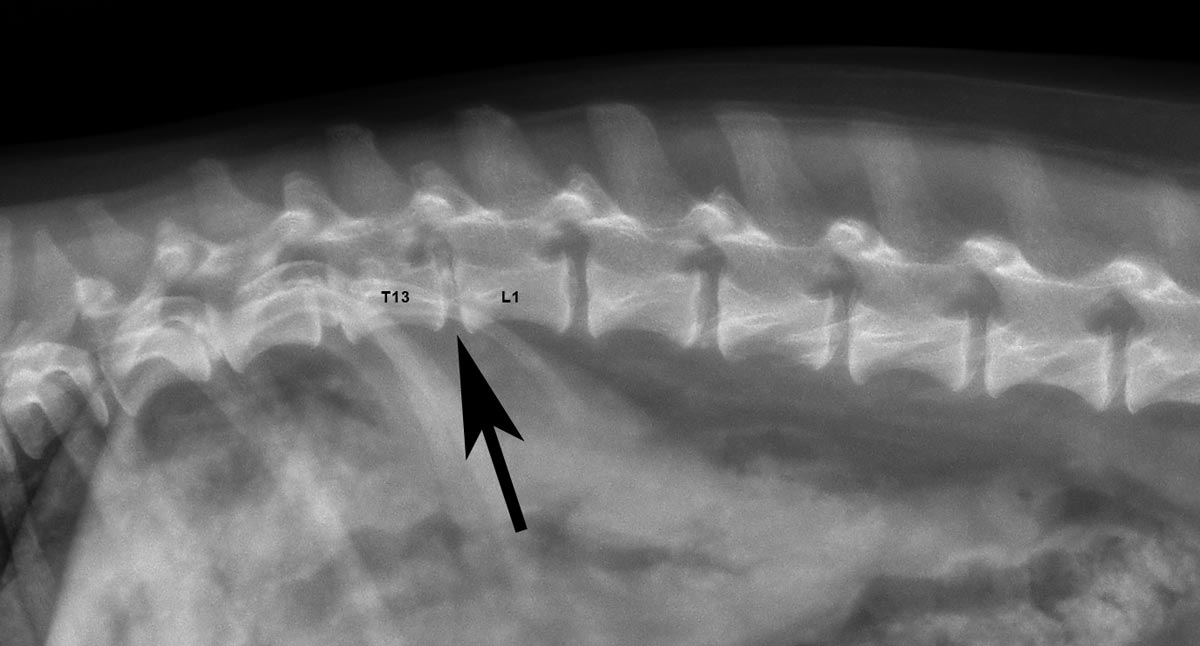
On December 13th, Hanz's family noticed he was less active, suspecting back pain. Radiographs revealed mineralization of three disc spaces, leading to a presumptive diagnosis of IVDD. Advanced imaging, such as MRI or CT, was recommended to fully assess the damage since soft tissue discs are not visible on radiographs.
Initially, anti-inflammatory medications provided some pain relief, but Hanz’s condition rapidly deteriorated to full paralysis and incontinence. Despite trying stronger medications with minimal improvement, Hanz’s family faced a heart-wrenching decision about his quality of life. Determined to explore every option, they sought a specialist for back surgery. An MRI confirmed severe, mineralized disc extrusion at T13-L1, causing near-complete spinal cord compression.
To alleviate the spinal cord constriction, Hanz underwent a hemilaminectomy, a surgical procedure to remove part of the vertebrae. By February 2024, Hanz was nearly walking normally again and has continued to recover, regaining his playful, spirited nature. Thanks to the dedication of his family and expert surgical care, Hanz's story is a testament to resilience and unwavering hope!
Sweet Abigail's story is a perfect reminder of the value of wellness care and the importance of following up on even mild abnormalities. While she had some chronic allergy issues, Abigail was otherwise thriving—eating well, drinking normally, and showing her usual sassy personality.
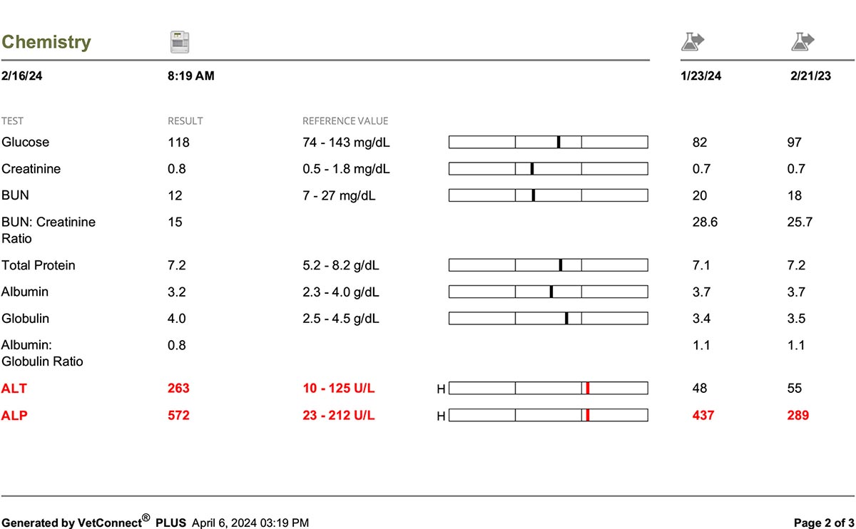
During her annual wellness exam in 2023, routine bloodwork began to reveal potential concerns. Specifically, two key enzymes used to assess liver and gallbladder health were flagged: ALT (alanine transaminase), which is associated with liver tissue, and ALP (alkaline phosphatase), which often reflects bile flow or gallbladder function but can also elevate due to medications, steroid use, or bone disease. Initially, Abigail’s ALP was only mildly elevated, a finding that can sometimes occur due to age, diet, or improper fasting before bloodwork. However, by the following year, her ALP had risen significantly, requiring intervention.
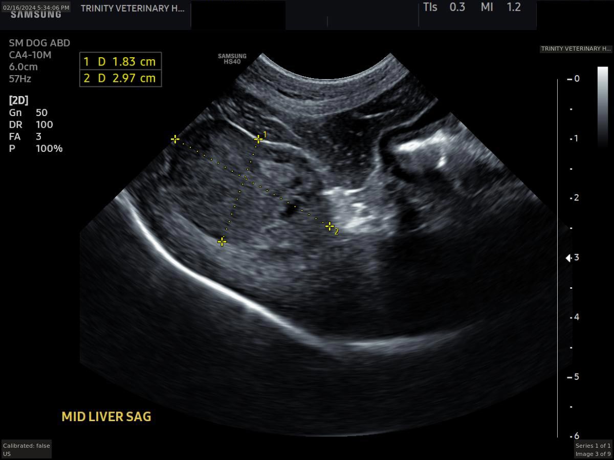
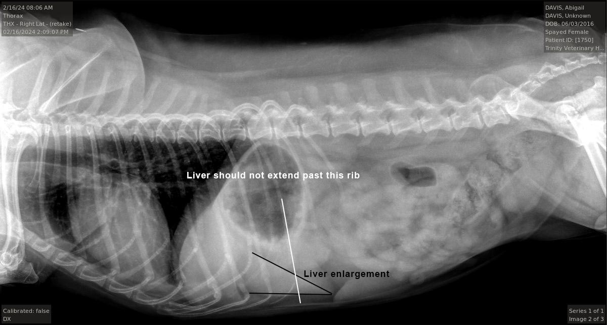
We started Abigail on medication and rechecked her levels three weeks later. Unfortunately, her ALP had climbed even higher, and her ALT was now elevated as well. This progression prompted further diagnostics. Radiographs were taken to assess her liver’s baseline structure and size, revealing mild enlargement as the liver lobe extended beyond the last rib. To investigate further, an ultrasound was performed to examine the liver tissue, gallbladder, bile duct, and other abdominal organs in greater detail. The ultrasound uncovered the source of the issue: a liver mass measuring 1.83 cm x 2.97 cm.
A fine-needle aspirate of the mass was attempted but did not yield a definitive diagnosis due to insufficient cells. Abigail was referred to a specialist for a CT scan and surgical consultation. Thankfully, the CT showed no evidence of additional tumors in her chest or abdomen, and the mass appeared surgically removable. Surgery was performed successfully, and Abigail is now recovering well. We are awaiting the pathology report and remain hopeful for a benign outcome.
Abigail’s case highlights why wellness care is so vital—it allows us to identify and address issues early, often before they become life-threatening. Abigail, no doubt, would agree!
Mochi has been a beloved patient at Trinity Veterinary Hospital since she was a puppy and a regular in our daycare program. Over the years, she occasionally experienced bouts of diarrhea, but in March 2023, something changed. This time, along with diarrhea, Mochi began whining and vomiting, and her abdomen was visibly painful. Given her history and current symptoms, an abdominal ultrasound was recommended.
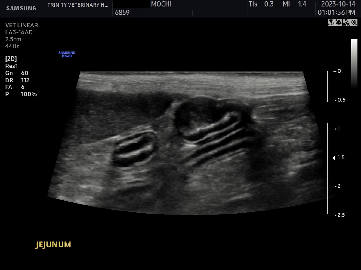
The ultrasound revealed an unexpected and unrelated finding. Within the layers of her intestine, there was a small, round area where the normal layering pattern was disrupted. This disruption raised concerns for either a tumor or a localized infection. Such an abnormality in a pet as young as Mochi is highly unusual. However, as this seemed incidental to her current gastrointestinal issues, Mochi was treated for her GI symptoms and quickly recovered.
Three months later, a follow-up ultrasound was performed, and the abnormal area in her intestine was still present, showing little to no change. To determine the nature of this finding, a surgical biopsy was performed. Tissue samples were sent for pathologic evaluation, and the results revealed that Mochi had MALT lymphoma, a very rare form of cancer.
Mochi was referred to an oncologist, and thanks to chemotherapy, she now has an excellent prognosis. Her case underscores the importance of thorough diagnostics and follow-up care, ensuring that even incidental findings are addressed with diligence. Mochi’s resilience and bright spirit continue to inspire us all!
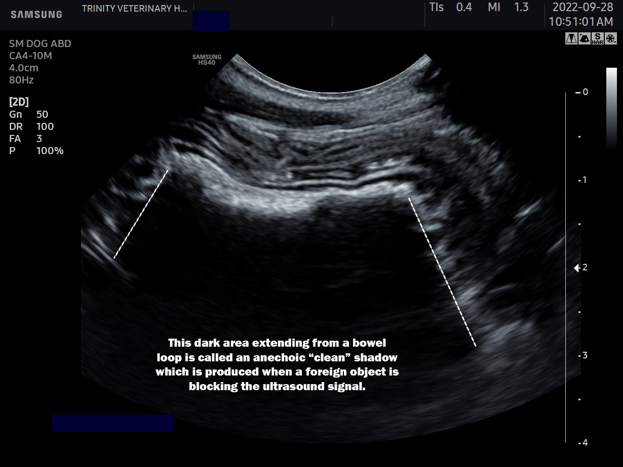
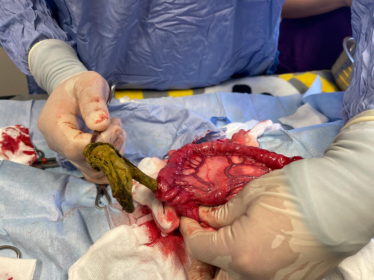
Blue came to Trinity Veterinary Hospital for persistent vomiting after receiving care at another hospital over the weekend. Upon examination, Dr. Williams noted that Blue was significantly dehydrated and showed mild abdominal pain during palpation. Radiographs were taken but revealed no definitive abnormalities. To investigate further, Blue was given barium, a contrast agent that enhances visibility on radiographs, to help identify a potential foreign body in the gastrointestinal tract. While most of the barium passed into the colon, some remained trapped in the small intestines. This finding was suspicious but not conclusive for a foreign body or surgical lesion.
Dr. Bonds performed an abdominal ultrasound, revealing thickened intestines and circular fluid accumulation—a sign that surgery might be necessary. The concern was for a potential foreign body or intussusception, a condition where one segment of the intestine telescopes into another.
Dr. Williams proceeded with an abdominal exploratory surgery and discovered a piece of stick, about 1.5 inches in diameter, and a sock lodged in Blue’s gastrointestinal tract. The stick was dangerously close to perforating the intestinal wall, which could have been fatal. Both the stick and the sock were carefully removed, preventing what could have been a life-threatening situation.
Blue made an excellent recovery, starting to eat the very next day. Her survival is a testament to the collaborative efforts of Dr. Williams and Dr. Bonds. Without timely intervention, Blue’s prognosis might have been dire, as intestinal perforation carries a survival rate of less than 30%. Blue’s case highlights the importance of thorough diagnostics and swift surgical intervention in emergencies.

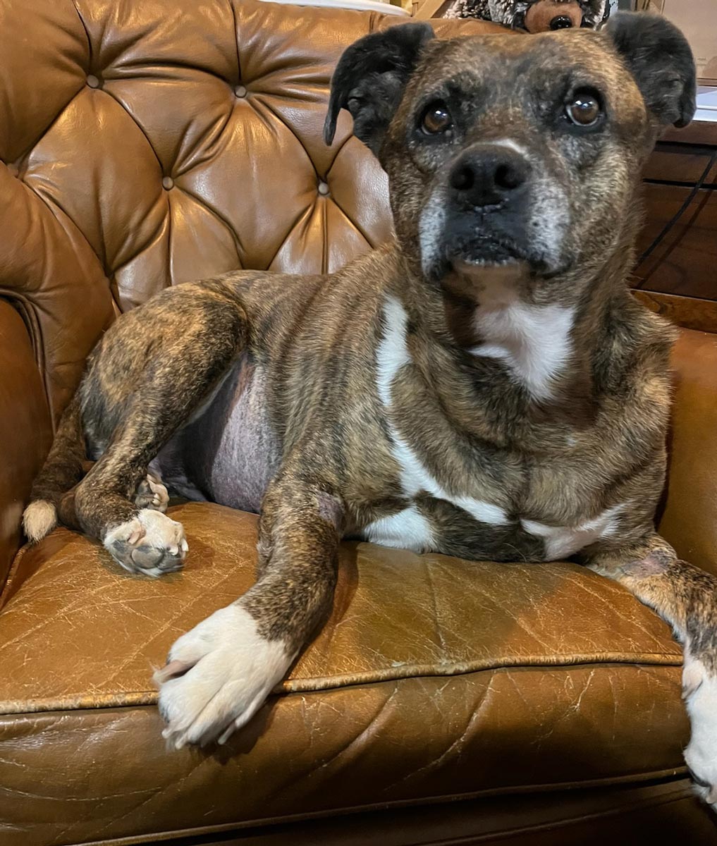 Jessie is one lucky girl! After being found on the streets, wary of anyone who tried to approach her, she was finally rescued by a loving family who has spoiled her ever since. Jessie was brought to Dr. Bonds for evaluation of a persistent cough, which the family had been told was related to a heart condition. During her examination, Dr. Bonds noticed that Jessie’s abdomen was distended. However, palpation was inconclusive due to Jessie’s tenseness during the exam.
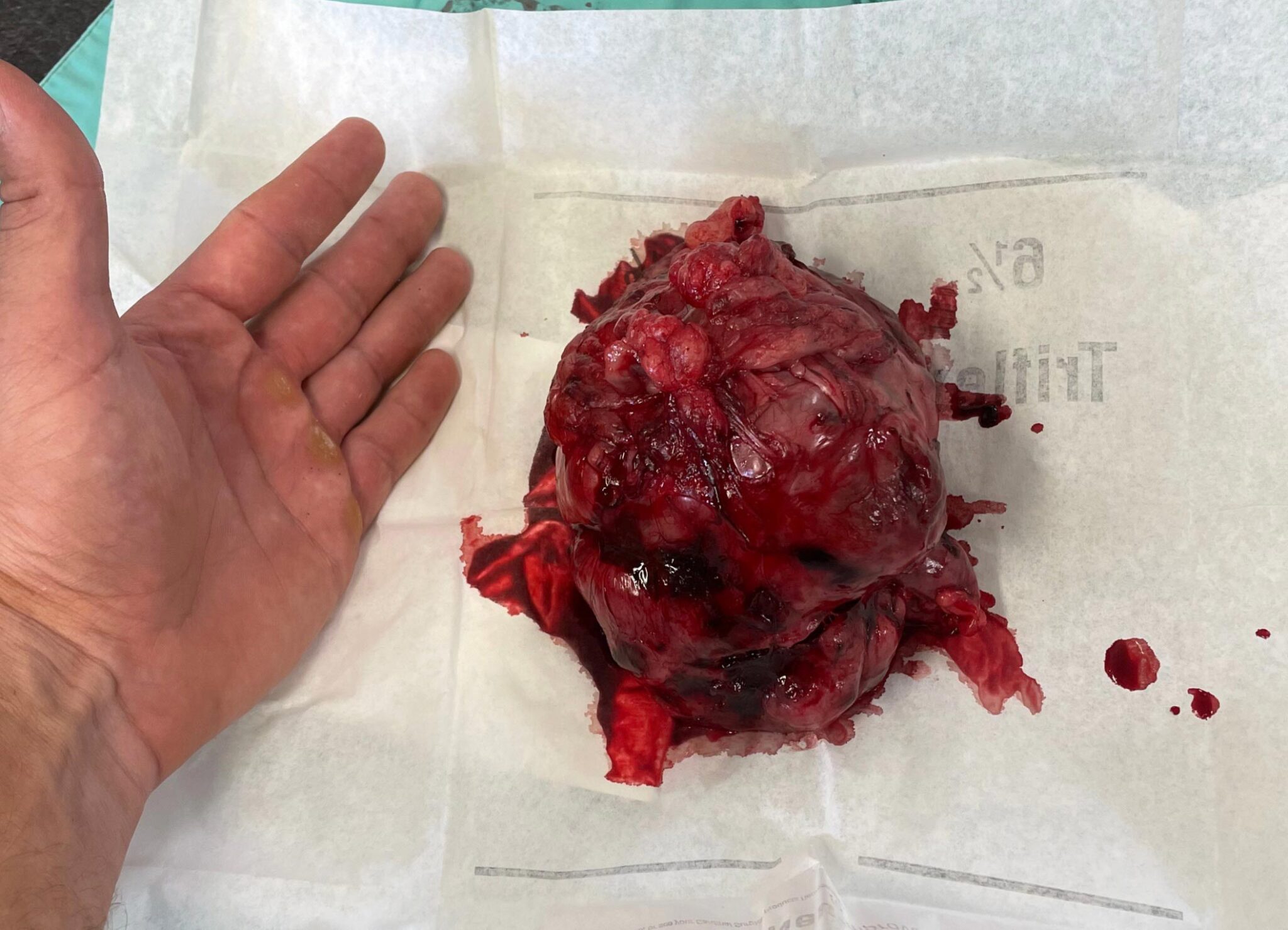
An abdominal ultrasound was performed, revealing a large mass of unknown origin. While the liver and spleen were initially suspected as potential sources, both appeared clear of any abnormalities. Faced with uncertainty, the family chose to pursue every option available. The next step was exploratory surgery to determine if the mass could be removed.
Dr. Williams performed the abdominal exploratory surgery and discovered that the mass originated from the cecum, a structure in dogs analogous to the appendix in humans. Thankfully, the mass was completely resected, and Jessie recovered well post-operatively.
While the ultrasound could not pinpoint the mass's origin, it provided critical information by ruling out visible signs of metastasis or involvement of other organs and lymph nodes. With the mass fully removed and no evidence of spread, Jessie’s prognosis is excellent. Her case is a wonderful example of how thorough diagnostics and timely surgical intervention can lead to a happy outcome!

 Brodie is an adventurous and energetic country dog who thrives on helping his owner with farm chores, riding tractors, feeding horses, and enjoying all things outdoors. Over the past six months, Brodie has faced a series of medical challenges, including a tick-borne infection, suspected leptospirosis, and a traumatic injury from getting tangled under an ATV (which leads to the next issue). His condition was a rollercoaster, with periods of improvement followed by setbacks.
At one point, Brodie developed a painless lump on his side, which remained stable for several months. However, this mass eventually ruptured, revealing an abscess. After several different therapies, it became clear that the infection was not responding to standard treatments, and Brodie needed surgery. Dr. Williams performed the procedure, and the source of the abscess was revealed: a single foxtail seed.
The abscess became life-threatening due to the presence of highly resistant bacteria. Brodie is now receiving daily injections of the only antibiotic effective against this strain, and he is currently responding well to treatment.
 Charlie is a 9-and-a-half-year-old Golden Retriever who is as sweet as they come, with nothing but love to give. He enjoys a normal life filled with family time and playdates with his doggie friends. However, one evening, Charlie suddenly began vomiting and showed signs of not feeling like himself. By the next morning, he had lost interest in both food and water and refused to leave his favorite spot in the yard. Concerned, his family rushed him to Trinity Veterinary Hospital, where Dr. Bonds conducted an examination. She noticed that his gum color was abnormal, which raised immediate concern for a potentially life-threatening condition.
Bloodwork revealed that nearly every value was abnormal, prompting Dr. Bonds to take X-rays, which showed a large amount of fluid in Charlie's abdomen—a strong indication of internal bleeding. The situation called for emergency exploratory surgery to determine whether a splenic tumor had ruptured or if another issue was causing the bleeding. After a lengthy three-hour surgery, the team successfully removed Charlie's spleen, and he made it through!
Thanks to the care and love of his family, Charlie is now recovering well and back to his happy, playful self.
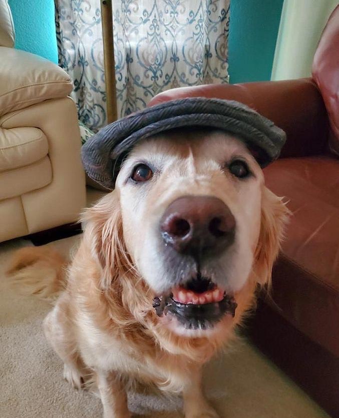
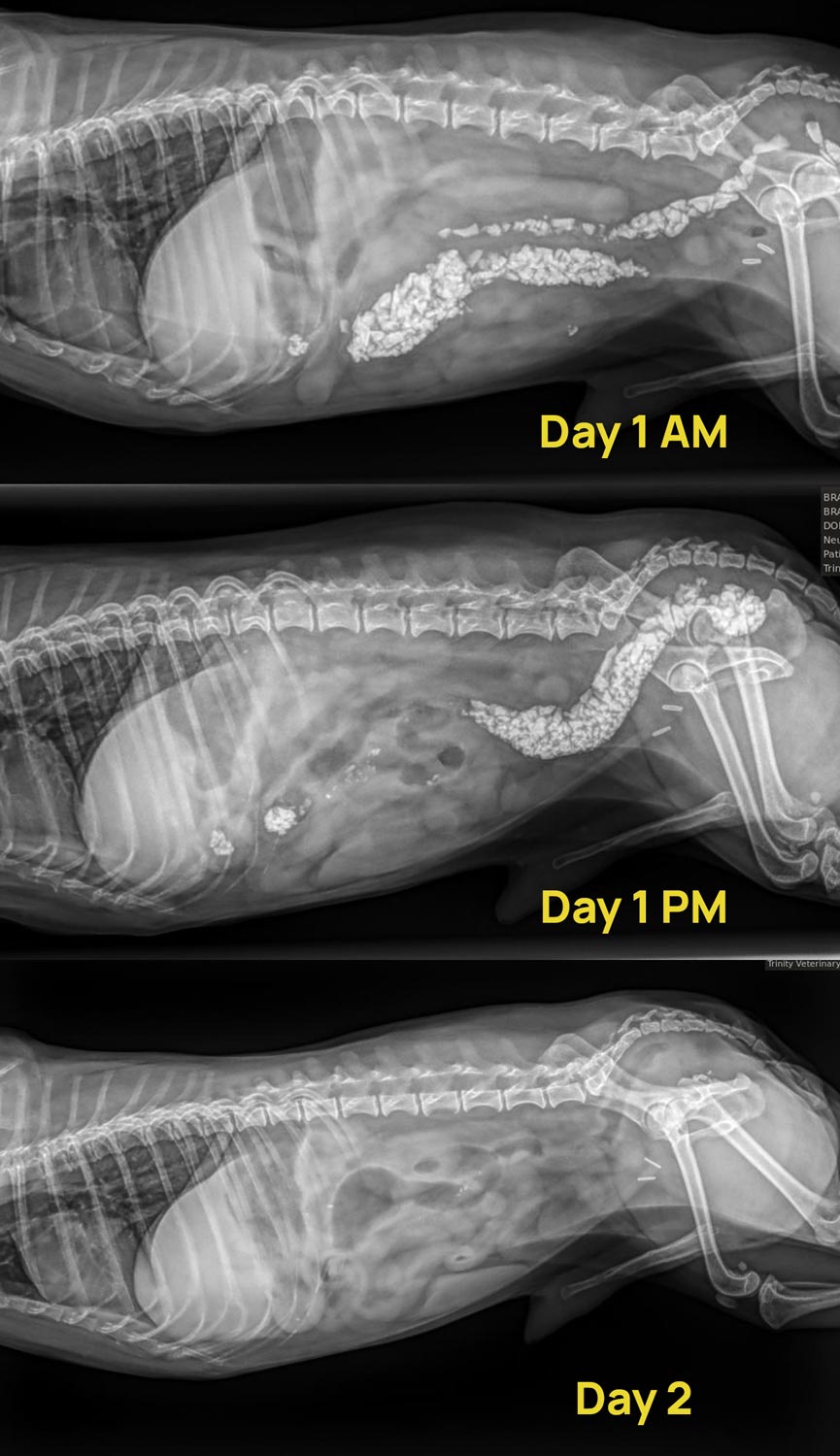 Do you ever wish you could peek into your dog’s brain, just for a few minutes to see why they do what they do? This is exactly what we would love to do with a precious baby who decided to eat enough small landscape rocks that they filled a large portion of his GI tract! Little Man is a sweet, 13 year old Yorkshire Terrier who has never craved rocks before but decided to eat enough that it threatened his intestines, and ultimately his life! He was vomiting rocks and had a large amount of bloody stool that contained, yep you guessed it – small, sharp little rocks. He was dehydrated, painful, and bloodwork showed his body was fighting infection stemming from his GI tract. He was immediately placed on IV fluids, injectable antibiotics, GI protectants, and pain medication. He would receive treatment for 24 hours and if the rocks were not moving through he would be headed for surgery. The treatment worked and the next morning he pooped out all of the rocks and radiographs confirmed what tiny bits remained would most likely work through on their own. He is at home eating and feeling back to his onery old self. We have talked to him at length about staying away from those pesky rocks – hopefully he listened and learned his lesson! We are so thankful he is doing well.
Kallie presented to Trinity Veterinary Hospital shortly after being hit by a car. Unfortunately, she was unable to walk when she arrived and was immediately carried to the treatment area. Her pain level was severe, so IV pain medications were started immediately following physical examination. There was an extensive open wound on the inner aspect of her right hind leg and the left hind leg had a large amount of bruising and swelling. Radiographs were taken of the chest and abdomen to look for evidence of internal bleeding and damage. No obvious abnormalities were seen. Radiographs of the rear limbs revealed that the left rear femur was broken in multiple pieces. After the radiographs, the large open wound on the inside of the right rear leg was cleaned and debrided (dead and dirty tissue was cut off). A tie over bandage was made with loops of suture so the bandage material could be placed over the wound like lacing a shoe to help keep the wound clean. The wound was cleaned, flushed and debrided as needed twice daily for 3 days. Supportive care and pain medications were continued throughout this time, with care taken to protect the fractured left hind leg as much as possible. After the wound was showing signs of healing and Kallie showed that she could support weight on her right hind leg, the difficult decision was made to proceed with amputation of the left rear limb. Amputation was needed because the severity of the fractures, along with Kallie’s other injuries, made repair very likely to result in failure and additional complications. Kallie stayed in the hospital with additional wound care and hospitalization for an additional 4 days after surgery. Kallie finally went home after over a week of in-hospital care at Trinity. Kallie’s owners continued at-home wound management under Dr. Williams’ advice for another 45 days before the open wound finally completely closed. Now, Kallie is running around happily on 3 legs like nothing ever happened. We are thrilled with the incredible progress Kallie has made!