Ultrasounds for Dogs: Uses & What to Expect

If your veterinarian has recommended an ultrasound for your dog, you probably have questions, and maybe a little anxiety. What exactly is a dog ultrasound? Does it hurt? What will it show? A veterinary ultrasound is a safe, non-invasive imaging tool that uses high-frequency sound waves to create real-time images of your dog’s internal organs and tissues. Unlike X-rays, which are best at evaluating bones and identifying masses, ultrasound lets your veterinarian see organ structure, blood flow, and soft tissue in detail, without any radiation. It is one of the most valuable diagnostic tools in veterinary medicine, and it gives your veterinary team information they simply cannot get any other way. Read on to learn when a dog ultrasound is recommended, what it can reveal, how to prepare, and exactly what to expect on the day of the procedure.
What Is a Dog Ultrasound Used For?
Veterinary ultrasound has a wide range of applications, and it is used in a variety of situations: from routine wellness evaluations to urgent diagnostic workups. Understanding why your veterinarian recommends an ultrasound can help you feel more confident going into the appointment.
Abdominal Ultrasound for Dogs
Abdominal ultrasound is among the most common uses of this technology in veterinary practice. It allows your veterinarian to evaluate organs including the liver, spleen, kidneys, bladder, stomach, intestines, adrenal glands, lymph nodes, and pancreas. This type of dog ultrasound is often recommended when a dog is experiencing symptoms such as vomiting, diarrhea, weight loss, increased drinking or urination, a distended abdomen, or suspected internal masses.
Abdominal ultrasound can identify abnormalities like masses or tumors, organ enlargement or atrophy, fluid in the abdomen, bladder stones, cysts, and changes in organ texture or structure that may indicate disease. It is also used to guide minimally invasive procedures like fine needle aspirates, which allow your veterinarian to collect tissue samples for analysis without the need for surgery.
Cardiac Ultrasound (Echocardiogram) for Dogs
A cardiac ultrasound, also known as an echocardiogram or echo, is used to evaluate the heart. It lets your veterinarian see the heart’s chambers, valves, walls, and surrounding structures in real time, as well as assess blood flow. This type of dog ultrasound is particularly useful for dogs that show signs of heart disease, such as exercise intolerance, coughing, fainting, or a heart murmur detected during a physical exam.
An echocardiogram can help diagnose conditions like dilated cardiomyopathy, mitral valve disease, pericardial effusion, and congenital heart defects. In dogs known to be at risk for heart disease such as Cavalier King Charles Spaniels, Doberman Pinschers, and Boxers, echocardiograms may be recommended as part of routine monitoring even before symptoms appear.
Pregnancy Ultrasound for Dogs
Ultrasound is the most reliable method for confirming pregnancy in dogs and monitoring fetal development. A canine pregnancy ultrasound can detect fetuses as early as 21 to 25 days after conception and allows your veterinarian to assess fetal heartbeats, count puppies, and evaluate placental health. It is a safe and stress-free alternative to other methods of pregnancy confirmation and gives breeders and dog owners important information as the due date approaches.
Other Common Uses of Dog Ultrasound
Beyond abdominal, cardiac, and pregnancy imaging, veterinary ultrasound is used to evaluate the prostate in intact male dogs, assess the reproductive tract in female dogs, examine the eyes and surrounding structures, investigate soft tissue injuries and musculoskeletal concerns, and guide minimally invasive procedures. Because ultrasound produces real-time images, it is also helpful in emergency situations such as when a dog presents with sudden collapse or abdominal distension, and can quickly rule in or out life-threatening conditions like internal bleeding.
How Is a Dog Ultrasound Performed?
One of the most common questions dog owners ask is whether an ultrasound will be uncomfortable for their pet. In most cases, a veterinary ultrasound is very well tolerated and does not require sedation. Here is what typically happens during the procedure.
Preparation Before the Ultrasound
For an abdominal dog ultrasound, your veterinarian may ask you to withhold food for several hours before the appointment. A full stomach can interfere with the image quality by trapping gas in the digestive tract. Your dog may also need to have the hair clipped from the area being examined, since fur can create air pockets that reduce image clarity. Warm ultrasound gel will be applied to the skin to help the transducer (the handheld device that emits and receives sound waves) make good contact with the surface.
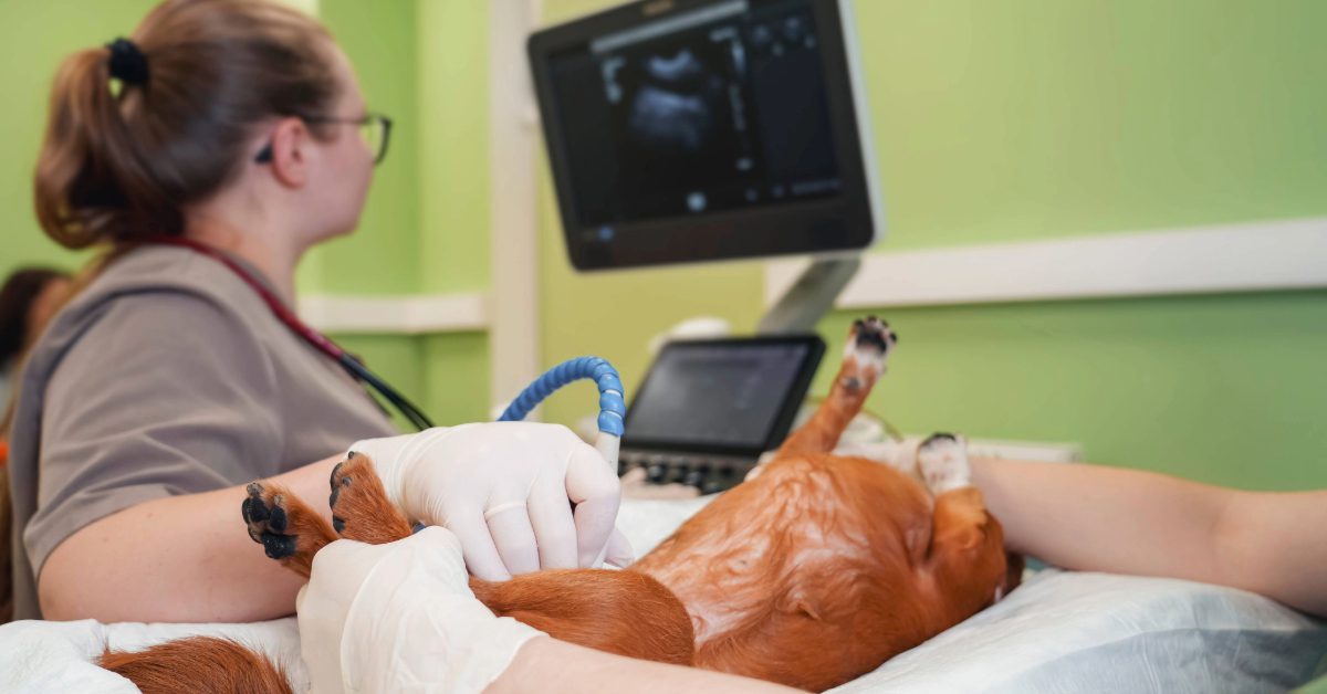
During the Procedure
Most dogs can remain awake and comfortable during an ultrasound. Your dog will typically be placed on their back or side on a padded table. A technician or veterinarian will hold the transducer against the skin and slowly move it across the area being examined while images appear on a monitor in real time. The procedure is painless, and many dogs remain quite relaxed throughout and some even fall asleep. If your dog is anxious or painful, a mild sedative may be recommended to help them stay still and comfortable. Your veterinary team will discuss this with you in advance if it is needed.
How Long Does a Dog Ultrasound Take?
The length of a veterinary ultrasound varies depending on the area being examined and what your veterinarian finds. A focused abdominal scan may take 20 to 30 minutes, while a full abdominal ultrasound or cardiac echo may take longer. If additional procedures like fine needle aspirates are performed at the same time, plan for the appointment to take a bit more time.
What Can a Dog Ultrasound Detect?
Dog owners are often surprised by the range of conditions that a veterinary ultrasound can identify. While no single diagnostic tool tells the whole story, ultrasound provides a level of detail that other imaging methods simply cannot match when it comes to soft tissue evaluation.
A dog abdominal ultrasound can detect masses, nodules, or abnormal growths in or around the organs; changes in organ size, shape, or echogenicity (texture); free fluid in the abdomen, which can indicate bleeding, infection, or organ rupture; bladder stones or polyps; kidney disease, including changes consistent with chronic kidney disease or acute injury; enlarged or abnormal lymph nodes; and evidence of splenic or hepatic disease.
Cardiac ultrasound for dogs can reveal the specific cause of a heart murmur, measure the size and function of the heart chambers, assess valve integrity, detect fluid around the heart, and guide decisions about cardiac medications. This level of detail is enormously helpful in tailoring treatment to your individual dog.
Ultrasound vs. X-Ray: What Is the Difference?
Many dog owners wonder whether their pet needs an X-ray or an ultrasound, and in many cases, the answer is both. These two imaging tools are complementary rather than interchangeable, and each has distinct strengths.
X-rays (radiographs) are excellent for evaluating bones, identifying the size and position of organs, detecting calcified structures like bladder stones, and assessing the lungs and chest. However, X-rays provide limited detail about what is happening inside the soft tissues of an organ.
Ultrasound, by contrast, excels at revealing the internal architecture of organs. It shows texture, fluid, vascularity, and movement in real time. It can also differentiate between solid masses and fluid-filled cysts, something X-rays cannot do. When your veterinarian recommends both imaging modalities, it is because they provide different and complementary information that together create a more complete picture of your dog’s health.
What Happens After a Dog Ultrasound?
After your dog’s ultrasound is complete, your veterinarian will review the images and discuss what was found. In some practices, results are available immediately; in others, images may be sent to a veterinary radiologist for specialist interpretation. If additional testing such as bloodwork, biopsy, or referral to a specialist is recommended based on the ultrasound findings, your veterinarian will explain why and walk you through the next steps.
Your dog will typically be able to go home the same day. If a fine needle aspirate or other procedure was performed during the ultrasound, there may be some mild soreness at the site, but recovery is generally quick and uncomplicated.

Answers Inside the Image
A dog ultrasound is one of the most powerful tools your veterinary team has for understanding what is happening inside your pet. It is safe, non-invasive, and provides real-time information that can guide faster, more accurate diagnoses and treatment decisions. If your veterinarian at Trinity Veterinary Hospital in Stillwater, OK has recommended an ultrasound for your dog, it is because they want to give your pet the most thorough evaluation possible. We are happy to walk you through the process, answer your questions, and make sure your dog is as comfortable as possible. Call us at (405) 533-0001 or book an appointment online to learn more about our diagnostic imaging services.
Recent Posts
About Us
For over a decade, Trinity Veterinary Hospital has provided compassionate, faith-based care to pets in our college town and beyond. We treat every pet like family, offering routine checkups, boarding, and advanced care from a board-certified internist.